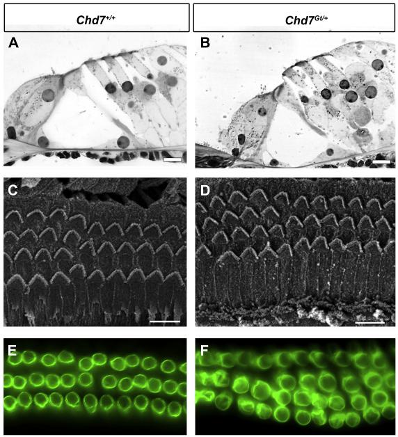
Figure 3. Cochlear anatomy and outer hair cell integrity is normal in Chd7Gt/+ mice.
Light microscopy (A-B), scanning electron microscopy (C-D) and Prestin epifluorescence (E-F) of the organ of Corti of wild type (A, C, E) and Chd7Gt/+ mice (B, D, F) at 6 weeks of age. (A-B) Histology for hair cells and non-sensory cells in the organ of Corti was similar in Chd7Gt/+ and wild type mice. (C-D) OHCs appear normal and the organization of surface morphology is similar between Chd7Gt/+ and wild type ears. In both wild type and Chd7Gt/+ mice, occasional stretches of 4 rows of OHCs are seen. (D-F) Prestin immunostaining of OHCs is unchanged in Chd7Gt/+ cochleae (D) compared to wild type (C) mice. Fourth row OHCs display Prestin staining that appears identical to that seen in the other three rows. Scale bars are 10 μm in A and B, and 20 μm in C and D.

