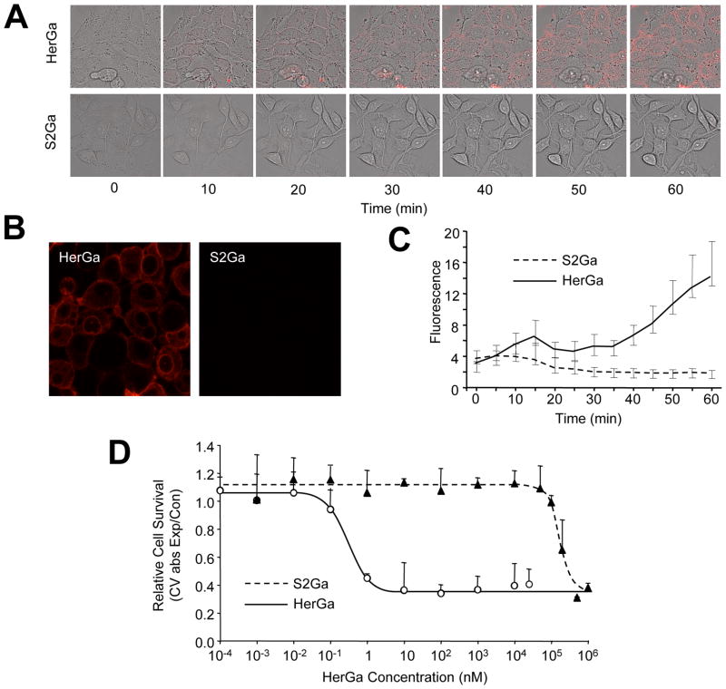
Fig. 1. HerPBK10 is required for enhanced internalization & cell death.
A, MDA-MB-435 cells were exposed to either HerGa or S2Ga (1 uM final corrole concentration) and imaged live by fluorescence confocal microscopy. Micrographs show fluorescence and brightfield overlays at key time points of uptake. B shows a comparison of fluorescence images acquired at 1h after HerGa or S2Ga uptake. C, Quantification of uptake in A. The cytosolic accumulation of fluorescence in each cell was quantified by selecting cytosolic regions and averaging fluorescence intensity using Image J. D, Cell death dose curve. MDA-MB-435 cells were incubated with HerGa or S2Ga at the indicated doses for 24h before cell survival was assayed by crystal violet (CV) stain. Cell survival is expressed as CV absorbance of each HerGa-treated sample normalized by mock (PBS) treated samples, or CV abs of experimental/control. Error bars represent 1 SD of triplicate treatments.

