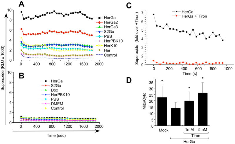
Fig. 5. Cytosolic HerGa induces superoxide generation.
MDA-MB-435 cells treated with 1 uM HerGa, S2Ga, HerPBK10, or PBS (mock) for 1h were analyzed for superoxide via chemiluminescence detection (see Methods). Graphs (A–C) displays relative luminescence unit (RLU) decay over time. A, Contribution of endosomolysis on superoxide generation. MDA-MB-435 cells were treated and assayed for chemiluminescence as described earlier. HerGa, HerGa-2, HerGa-3, and S2Ga were added to cells at 1 uM corrole concentration. Individual components were added at the equivalent protein concentration to each respective complex. B, Lack of superoxide generation in a cell-free system. HerGa (1 uM) and equivalent concentrations of S2Ga, HerPBK10, or PBS were added directly to superoxide anion assay medium and luminol oxidation was measured as described in the Methods. C, Reduction of HerGa-mediated superoxide generation in MDA-MB-435 cells by the superoxide scavenger, Tiron. Cells were incubated with 1 mM Tiron for 1h before treatment with 1 uM HerGa for 24h, followed by superoxide measurement as described earlier. D, Effect of Tiron on HerGa-mediated ΔΨ (m) disruption. Cells were incubated with the indicated concentrations of Tiron before treatment with 1 uM HerGa for 24h, followed by measurement of TMRM uptake as described earlier. *, P<0.02 compared to HerGa alone, as determined by two-tailed unpaired t test.

