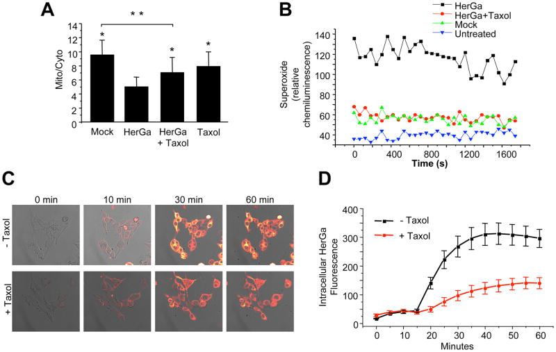
Fig. 7. Microtubule stabilization abrogates events downstream of HerGa uptake.
MDA-MB-435 cells were incubated in media containing 5 uM taxol for 15 min before exposure to HerGa (see Methods). At 24h after exposure, cells received (A) 20nM TMRM and were monitored for mitochondrial TMRM accumulation as described previously; or (B) luminol and assayed for superoxide levels as described in the Methods. *, P<0.0001 compared to HerGa treatment. **, P<0.0001. Statistical significances determined by two-tailed unpaired t-tests. C–D, Effect of taxol on HerGa uptake. C, Confocal images were acquired of live MDA-MB-435 cells after treatment with 5uM HerGa (−/+ pretreatment with 5 uM taxol; see Methods). D, Cytosolic fluorescence from the same cell region per time point was measured and average fluorescence variations plotted over time, comparing HerGa −/+ taxol.

