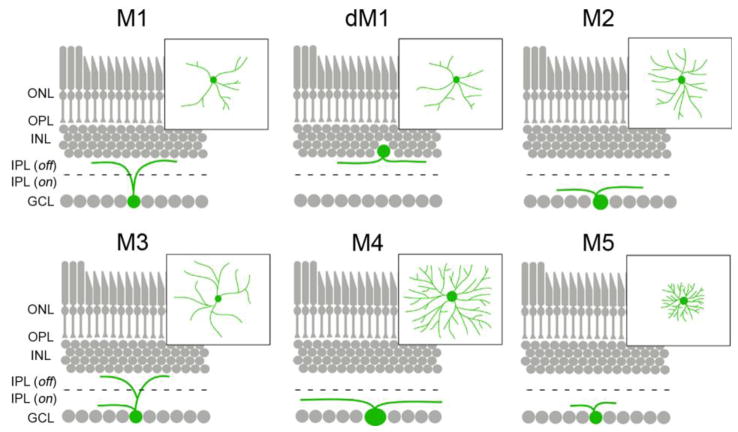
Figure 2.
Morphological differences between classes of ipRGCs. Stratification of the dendrites of each of the 5 classes of ipRGC (and displaced M1 ipRGCs [dM1]) are depicted in retinal cross-sections. Insets show the morphology of dendritic arborizations for each class as seen in retinal whole-mount preparations. Black dashed line indicates the division of the inner plexiform layer into ON and OFF divisions of the IPL. ONL – outer nuclear layer; OPL – outer plexiform layer; INL – inner nuclear layer, IPL(off) – OFF division of the inner plexiform layer; IPL(on) – ON division of the inner plexiform layer; GCL– ganglion cell layer.

