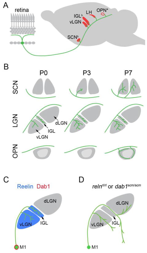
Figure 4.
Development of central projections of M1 ipRGCs, A. 5 nuclei (red) receive dense projections from M1 ipRGCs (green): suprachiasmatic nucleus (SCN), ventral lateral geniculate nucleus (vLGN); intergeniculate nucleus (IGL), lateral habenula (LH) and the olivary pretectal nucleus (OPN). ‘b’ denotes those nuclei that receive binocular input from M1 ipRGCs. ‘*’ denotes that only the outer ‘shell’ of the OPN is innervated by M1 ipRGCs. B. Development of M1 ipRGC innervation to the SCN, lateral geniculate nucleus (LGN, which is composed of the vLGN, IGL and dorsal LGN [dLGN]), and OPN at postnatal day 0,3 and 7 (P0, P3, P7 respectively) in mice. vLGN and IGL are innervated by M1 ipRGCs by P0, while SCN is innervated by P3 (although only the contralateral SCN is innervated by M1 ipRGC axons at this early age), and the ‘shell’ of the OPN is innervated by P7. C. Reelin (blue) is expressed in the ventral lateral geniculate nucleus (vLGN) and intergeniculate nucleus (IGL). M1 ipRGCs (M1) express disabled-1 (Dab1; red), an intracellular component of the reelin signaling pathway. D. M1 ipRGC axons are mistargeted in spontaneously generated mouse mutants lacking either Reelin (relnrl/rl) or Dab1 (dab1scm/scm).

