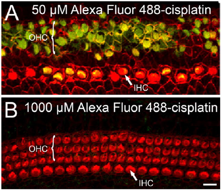
Fig. 5.

Accumulation of cisplatin in hair cells was greater following 48 h exposure to a low-versus high concentration. (A and B) Representative confocal photomicrographs show the intracellular accumulation of cisplatin conjugated to a fluorescent probe, Alexa Fluor 488 (green), in OHCs and IHCs. (A) At a concentration of 50 μM, Alexa Fluor 488-cisplatin accumulated preferentially in the OHCs. Consistent with this cisplatin accumulation, the OHCs showed significant structural damage. (B) Following treatment at 1000 μM, hair cells were intact and showed little accumulation of Alexa Fluor 488-cisplatin. Scale bar shown in panel B represents 10 μm.
