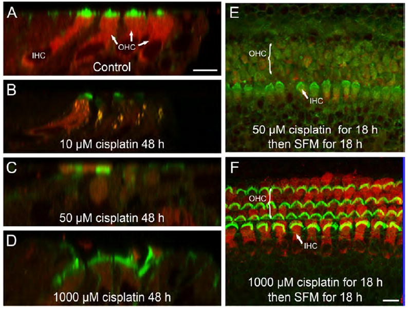
Fig. 6.

Stereocilia damage was induced by low, but not high concentrations of cisplatin. (A-D) Representative confocal micrographs show the uptake of FM1-43 (red) in IHCs and OHCs in control and cisplatin-treated (10, 50 and 1000 μM) cultures. Hair cell stereocilia are labeled green. (E and F) FM1-43 uptake was visualized after 18 h of cisplatin treatment (50 or 1000 μM) followed by 18 h of exposure to normal serum-free medium (SFM). Unlike hair cells exposed to 50 μM cisplatin (E), those treated with a high concentration of cisplatin (F) were able to uptake FM1-43 after the ‘recovery period’, which indicated that the stereocilia were not permanently damaged. Scale bars shown in panels A and F each represent 10 μm.
