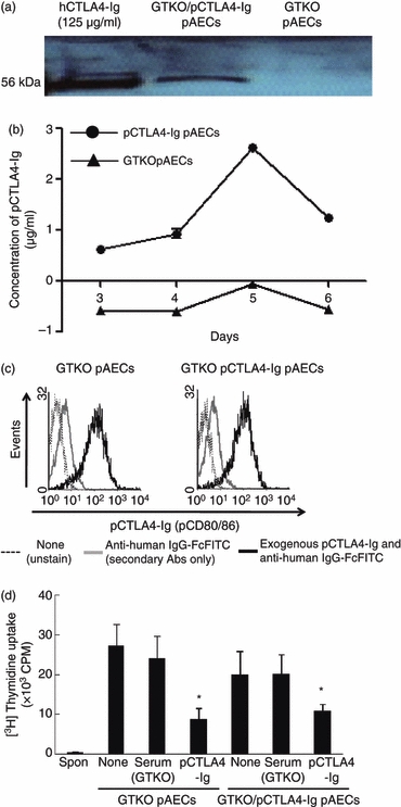
Figure 7.

Absence of a direct suppressive effect on the human (h) CD4+ T-cell response to porcine aortic endothelial cells (AECs) expressing porcine cytotoxic T-lymphocyte antigen 4 immunoglobulin (pCTLA4-Ig) was associated with weak of production of soluble pCTLA4-Ig during culture. (a) Detection of pCTLA4-Ig protein in pAECs from α1,3-galactosyltransferase gene-knockout (GTKO)/pCTLA4-Ig pigs by Western blot analysis. A specifically positive band (56 000 molecular weight) was observed in lysates of GTKO/pCTLA4-Ig pAECs, but not in lysates of GTKO pAECs. Purified hCTLA4-Ig (125 μg/ml) was loaded as a positive control. (b) Detection by ELISA of pCTLA4-Ig in the supernatant from cultured pAECs expressing pCTLA4-Ig. GTKO/pCTLA4-Ig and GTKO pAECs (1 × 10⁶ cells) were cultured in a 75T flask from 3 days to 6 days without changing the culture medium. Approximately 500 μl supernatant was collected from the cultured cells at 3, 4, 5 and 6 days after culture. Levels of pCTLA4-Ig were measured by ELISA with serial dilution. (c) Detection by flow cytometry of pCTLA4-Ig bound to pAECs. GTKO/pCTLA4-Ig and GTKO pAECs were cultured in 75T flasks until confluence. The pAECs were harvested and tested to determine whether soluble pCTLA4-Ig produced by GTKO/pCTLA4-Ig pAECs bound to these pAECs in an autocrine fashion during culture. Porcine AECs were stained with/without FITC-conjugated anti-human IgG-Fc antibodies, and fluorescence intensity in pAECs was compared between unstained (solid line) and stained (with FITC-conjugated anti-human IgG-Fc antibodies) (grey line), and between GTKO pAECs and GTKO/pCTLA4-Ig pAECs. The expression of pB7 molecules on GTKO and GTKO/pCTLA4-Ig pAECs was also compared by staining with pCTLA4-Ig (500 μg/ml) followed by FITC-conjugated anti-human IgG-Fc antibodies (solid thick line). Results are representative of three independent experiments. (d) The direct effect of pAECs expressing pCTLA4-Ig on the hCD4+ T-cell xenogeneic response was investigated by mixed lymphocyte reaction. The hCD4+ T cells were co-cultured with GTKO and GTKO/pCTLA4-Ig pAECs untreated or treated with either GTKO serum or soluble pCTLA4-Ig (500 μg/ml) (n = 5). Results were compared with hCD4+ T-cell responses to GTKO pAECs and GTKO serum-treated stimulators. (*P< 0·05 versus GTKO serum-treated).
