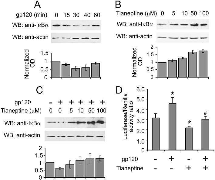
Figure 4.

Tianeptine interferes with NF-κB signalling by stabilizing IκB-α in astroglial cells. (A) Time-course analysis of IκB-α protein levels in response to gp120 stimulation. The astroglial cells (seeded 1.5 × 104 cm−2 cells 24 h before the treatments) were stimulated with 10 nM gp120 and lysed in RIPA buffer at indicated time points. Lysates were processed and 5 µg of total protein lysates were loaded on the 10% SDS-PAGE gel and subsequently analysed by Western blotting. The blot is representative of three independent experiments. The graph represents the normalized mean OD of IκB-α signal divided by mean OD of actin signal from three independent blots ± SEM. (B) Tianeptine stabilizes IκB-α in the absence of gp120 stimulation. The cells seeded as in (A) were treated for 1 h 40 min with different concentrations of tianeptine and processed as in (A) for Western blotting. The graph represents the normalized OD of IκB-α signal from analysed as in (A). (C) Tianeptine inhibits the degradation of IkBa induced by gp120. The astroglial cells were seeded as in (A) and stimulated with gp120 for 35 min. Tianeptine at different doses was added 1 h before the administration of gp120. The cells were processed for Western blotting as in (A). The graph shows the normalized OD of IκB-α signal analysed as in (A) from 3 independent experiments. (D) NF-κB transcriptional activity is suppressed by tianeptine. The astroglial cells were transfected with pNfkB-Luc and pRL reporter plasmids. Twenty hours post-transfection, the cells were stimulated with 10 nM gp120 and/or tianeptine (50 µM) for 5 h or left untreated. Each treatment was performed six times. Luciferase activity and internal control renilla activity are the mean of six experimental points. Data represent the mean ± SEM of a representative experiment, which was performed three times. *P < 0.05 when compared with control; #P < 0.05 tianeptine and gp120 versus gp120 alone-treated cells.
