
Figure 2.
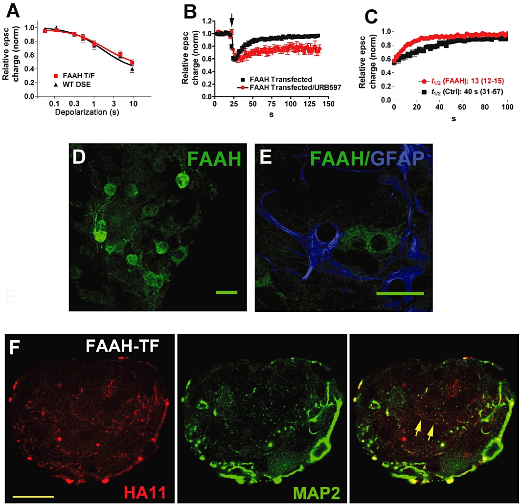
Overexpression of FAAH with endogenous MGL also shortens DSE duration. (A) ‘Dose’–response for DSE of FAAH-transfected neurons (FAAH T/F) using a range of depolarizations from 50 ms to 10 s. The wild-type DSE dose–response is shown for comparison. (B) Averaged DSE time courses for neurons transfected with FAAH and FAAH transfected neurons subsequently treated with URB597 (250 nM). (C) DSE recovery time courses for FAAH-transfected and control cells. t1/2-values with 95% CIs are listed below the curves. (D) Micrograph shows FAAH staining in autaptic hippocampal cultures. Scale bar = 25 µm. (E) Micrograph shows FAAH staining does not colocalize with the astrocyte cell marker GFAP. Scale bar = 25 µm. (F) Micrograph shows that FAAH is expressed in neurons transfected with FAAH, and that it is expressed in processes that are labelled and unlabelled with dendritic marker MAP2. Left panel shows HA11 staining against the HA-tagged FAAH protein. Centre panel shows dendritic marker MAP2. Right panel shows the composite of the left and centre panels, with overlap in yellow. Scale bar = 15 µm.