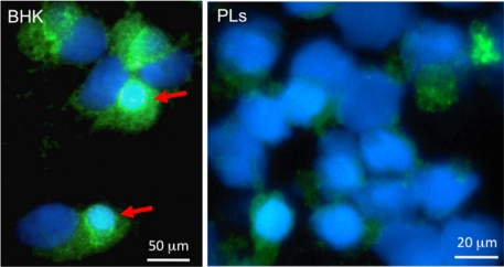
Figure 4.
Immunofluorescence microscopy of baby hamster kidney cells (BHK, left) and Xenopus PLs infected in vitro for 2 days with FV3 (0.3 MOI). Cells were cytocentrifuged on microscope slides, fixed with formaldehyde, permeabilized with ethanol, incubated with a rabbit anti-53R and FITC-conjugated donkey anti-rabbit Abs (Green); then stained with the DNA dye Hoechst-33258 (Blue) mounted in anti-fade medium and visualized with a Leica DMIRB inverted fluorescence microscope. Note the large viral assembly sites in BHK cells that contain large amount of viral DNA stained Hoechst-33258 and anti-53R Ab (arrows). In contrast, anti-53R staining is weaker in PLs, and no assembly sites are detected.

