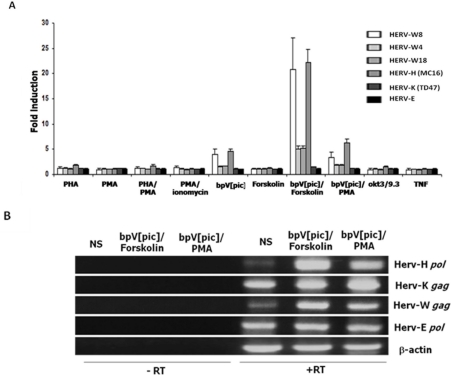
Figure 1.
Increased expression and promoter activity of different HERVs after T-cell activation. (A) Reporter plasmids carrying the complete 5′LTR of different HERVs were transfected into Jurkat T-cells. At 24 h post-transfection, cells were stimulated with PHA, PMA, PHA/PMA, PMA/ionomycin, bpV[pic], Forskolin, bpV[pic]/Forskolin, bpV[pic]/PMA, OKT3(anti-CD3)/9.3 (anti-CD28) and TNFα. After stimulation (8 h), cells were lysed and measured for luciferase activity. Results are shown as fold induction relative to luciferase activity of untreated cells and are the mean of three independently treated cell samples. (B) Total RNA was extracted from cells treated or not with bpV[pic]/Forskolin or bpV[pic]/PMA and RT-PCR was performed for the detection of the following transcripts: the gag gene of HERV-K and HERV-W, the pol gene of HERV-H and HERV-E and β-actin. Expression levels were compared with the RNA from non-stimulated Jurkat cells (NS). Controls consisted of RT-PCR reactions conducted in the presence of RNA with no RT enzyme.

