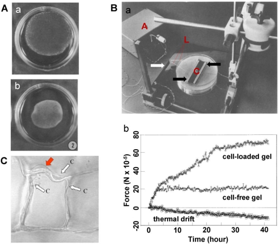
Figure 2.
The cellular contractile forces sensed using a CPCG model. A. (a) Collagen gel contracts and exhibits a decrease in size; (b) the collagen gel further contracts, and its size is further reduced (adapted with permission from Figure 2 in [23]). B. (a) An experimental set-up for culture force monitor. Microporous polyethylene bars (indicated by the black arrows) are attached to a collagen gel and float in culture medium. The strain gauge beam is marked with a white arrow. The beam and a bar are connected using an A-shape frame (L) made from stainless steel suture wire. The amplifier (A) is also shown. (b) Cell forces change with time (adapted with permission from Figures 1–3 in [19]). C. Within a collagen-GAG foam-like gel, an individual dermal fibroblast (red arrow) elongated and deformed several surrounding struts (white arrows) in the scaffold (adapted with permission from Figure 1 in [26]).

