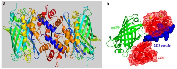
Figure 1.
Schematic representation of Case16 structure A. (a) Case16 crystallized as a monomer in the asymmetric unit (in rainbow coloring mode: N-terminus blue, C-terminus red). The crystallographic contact between C-terminal lobes of CaM of the two Case16 molecules is a result of a packing interaction rather than a binding interaction. (b) Domain representation of Case16 structure A. M13-peptide is colored blue, cpGFP is green and N- and C-terminal lobes of CaM are shown red. Ca2+ ions are shown as yellow spheres.

