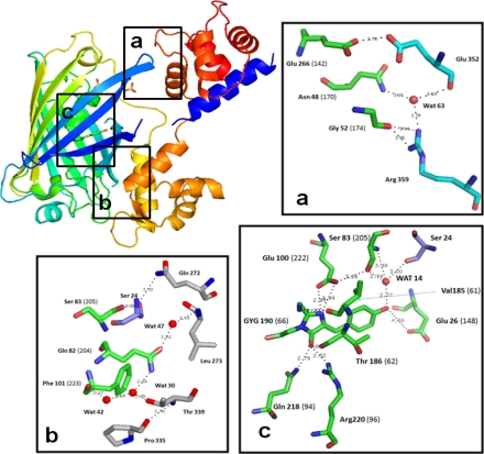
Figure 2.
Ribbon diagram and key residues of the chromophore environment of Case16 structure A (colored in rainbow mode: N-terminus blue, C-terminus red). Insert “a” shows the loose contact of cpGFP with the C-terminal CaM lobe. Insert “b” shows the tight contact with the N-terminal lobe which is dominated by hydrophobic interactions. Insert “c” shows the local environment of cpGFP chromophore. Residue numbers indicated in brackets refer to wtGFP numbering. Residues with carbon atoms in green belong to cpGFP domain, residues with carbon atoms in cyan belong to CaM-domain and residues with carbon atoms in blue belong to M13-peptide.

