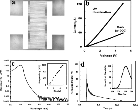
Figure 16.
(a) Optical microscope picture of a Mg0.34Zn0.66O UV detector with MSM structure. (b) I-V curves show dark current and photocurrent under 308 nm, 0.1 μW UV light illumination. (c) Spectral response of a Mg0.34Zn0.66O UV detector biased at 5 V. (d) Temporal response of Mg0.34Zn0.66O UV detectors excited by nitrogen gas laser pulses (337.1 nm, <4 ns) [90].

