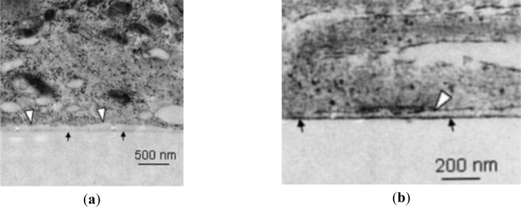
Figure 7.
TEM images showing examples of cell-substrate clefts—from [75]. Cells have been fixed and sectioned using a focussed ion beam: (a) A platinum substrate (the surface marked with black arrows) was coated with laminin-111 prior to adhesion of chicken embryo neurons. The cleft is between the adhered cell membrane (marked by white arrows) and the platinum surface and was measured to be 27–108 nm; (b) L1 Ig6 (the sixth immunoglobulin domain of cell adhesion molecule L1 and known to promote neurite extension) has a lower molecular weight (8 kDa) and is a smaller molecule than laminin-111 (∼800 kDa). Smaller molecules generally result in smaller clefts, as illustrated here by the cleft of 26–79 nm.

