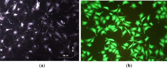
Figure 6.
(a) The observation of articular chondrocyte morphology during cell culture period using a dark field microscope; and (b) the observation of cell viability after 3 day perfusion cell culture using the Live/Dead® fluorescent dye and fluorescent microscope (Green and red dots represent live and dead cells, respectively).

