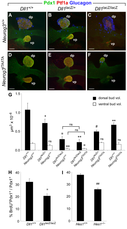
Fig. 5.
Ptf1a-mediated Dll1 expression is required for MPC proliferation. (A-F) Image stack projections of E10.5 embryos from crosses of Dll1lacZ/+Neurog3tTA/+ double-heterozygote animals, whole-mount stained for Pdx1, Ptf1a and glucagon. Note that dorsal and ventral bud size is equally reduced in Dll1lacZ/lacZNeurog3+/+ and Dll1lacZ/lacZNeurog3tTA/tTA embryos. dp, dorsal pancreas; vp, ventral pancreas. Scale bars: 50 μm. (G) Quantification of dorsal and ventral bud volume in wild-type and mutant embryos of the indicated genotypes based on Pdx1/Ptf1a double whole-mount stained and confocally scanned embryos. (H,I) Quantification of BrdU incorporation in E10.5 Pdx1+ MPCs in Dll1 and Hes1 mutant dorsal buds, respectively. Data in G-I are represented as mean ± s.d. ##, P<0.0002; 
 , P<0.0005; #, P<0.002;
  , P<0.005; **, P<0.01; *, P<0.05; by Student’s t-test (compared with wild type unless otherwise indicated); n=2-4. ns, not significant.

