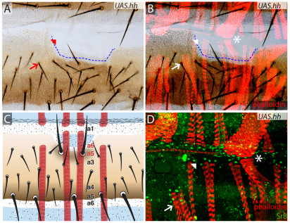
Fig. 6.
UAS.hh clones change the identities of the epidermal cells as well as the positions of muscle attachments. (A-C) UAS.hh-expressing clone (boundary estimated with a blue dashed line) located in a2/a3 area, transforms its cells towards P identity and as Hh spreads from the clone into surrounding A territory it changes the identity and orientation of neighbouring cells, replacing a2/a3 and a3 with a6-a4 (Struhl et al., 1997b) as shown schematically in C. The muscles underlying the transformed cuticle do not contact the newly formed a4-a6 cuticle and instead attach to a1 cuticle that is still present (asterisk). Note that muscles from the preceding segment attach correctly even in the neighbourhood of a UAS.hh clone, presumably because a1 is not changed by the clone. (D) Detail showing the muscle attachments and apparently normal tendon cells (green, marked with anti-SrB). Asterisks, arrows and arrowheads indicate the corresponding points in the three figures.

