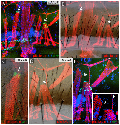
Fig. 7.
UAS.sr clones attract muscles and divert them from their usual points of attachment. (A,B) UAS.srB clones (marked with GFP, green) attract the muscles (stained with phalloidin, red). All tendon nuclei are marked with anti-SrB antibody (blue). Some muscles attach to ectopic sites where sr is expressed (arrows). (C-E′) Details of clones shown in A and B. Note the altered cuticle in one sr-expressing clone (arrow, C) while another clone can make maimed bristles (below arrow, D). (E) The same region stained for anti-SrB antibody (blue) and GFP for UAS.srB (green); (E′) A detail of E lacking the green channel showing that both the endogenous and ectopic tendons express sr at similar levels. It appears that myotubes ignore their usual attachment sites and prefer to form ectopic contacts with the clones, either because the ectopic sr expression starts earlier or is more persistent; if the latter, this could be due to autoregulation of sr (Vorbruggen and Jackle, 1997). Asterisk in E,E′ indicates the same location.

