Figure 5.
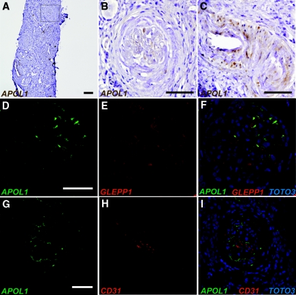
APOL1 redistribution is consistent between FSGS and HIVAN samples. Immunoperoxidase (A–C) and confocal immunofluorescent images (D–I) of kidney biopsy specimens from patients with HIVAN stained using anti-APOL1 antibody. (A) Reduced APOL1 staining in glomeruli and cortical tubules compared with the normal kidney. (B) Selected glomerulus from (A) shows diminished APOL1 staining (arrowhead). (C) In contrast to the normal kidney, APOL1 also appears in vessel wall of the renal arterioles. (D & F, green) APOL1 signal is diminished in the glomerulus, but remains colocalized with GLEPP1 expression (E & F, red), which is also diminished in HIVAN. (H & J, green) Glomerular APOL1 signal does not overlap with CD31 staining (I & J, red), which identifies glomerular capillary endothelium. (F & I, blue) Nuclei were visualized with TOTO-3 staining. Scale bars: 100 μm (A), 50 μm (B–I).

