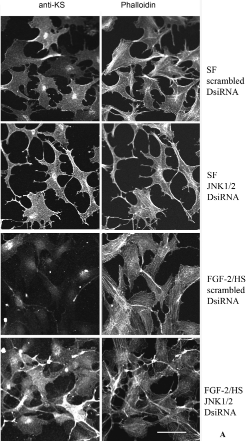
Figure 7.
TGF-β1– and FGF-2/HS–induced decrease in the cell surface–associated KS is occluded by JNK1/2 transfection of keratocytes. A portion of DsiRNA transfected keratocytes, from the experiments described in Figure 5, activated with 40 ng/mL FGF-2 and 5 μg/mL HS (A), or with 2 ng/mL of TGF-β1 (B), were plated for immunocytochemical analyses. Double fluorescence staining was performed using mouse anti-KS antibody followed by Alexa Fluor 488-anti-mouse IgG antibodies and Alexa Fluor 546-phalloidin. Bar, 50 μm.

