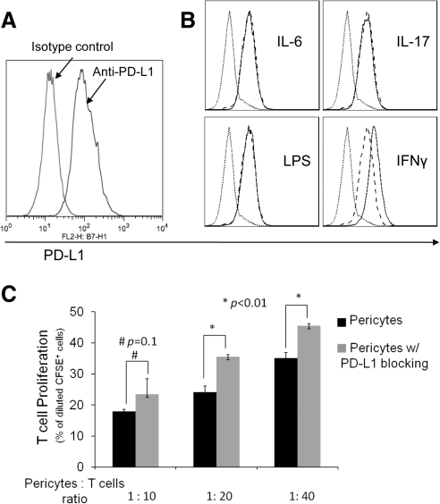
Figure 4.
Role of PD-L1 in RPC-mediated T cell inhibition. (A) RPCs express PD-L1. RPCs (0.5 × 10⁶) were stained with 5 μg/mL PE-labeled anti-PD-L1 IgG or matched isotype control, then analyzed by flow cytometry. (B) PD-L1 on RPCs is upregulated by IFN-γ. RPCs (1 × 10⁵/mL) were incubated without and with IL-6, IL-17, LPS, or IFN-γ for 24 hours. After washing, levels of PD-L1 were assessed by flow cytometry (dotted line, isotype control; dashed line, without treatment; thin line, with stimuli). (C) Blocking PD-L1 activity reduces the efficacy of RPCs in inhibiting T cells. The same CFSE-based T cell proliferation assays were performed using activated T cells and different numbers of RPCs in the presence of 5 μg/mL anti-PD-L1 IgG or isotype controls (azide free).

