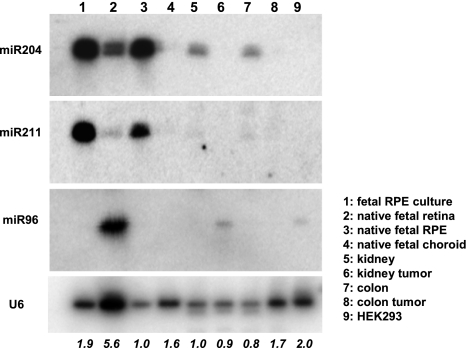
Figure 3.
Northern blot expression of miR-204, miR-211, and miR-96 was analyzed by Northern blots with LNA-spiked and 32P-labeled oligonucleotides as probes. RNA of human fetal retina (lane 2), RPE (lane 3), and choroid (lane 4) were pooled from 3 individual donors. RNA of cultured RPE (lane 1) was obtained from passage 1 of hfRPE primary cultures. Total RNAs of normal and malignant adult human kidney (lanes 5 and 6) and colon (lanes 7 and 8) were purchased from Ambion. Total RNA (10 μg) from each preparation was loaded onto a 15% acrylamide gel. Native (lane 3) as well as cultured (lane 1) RPE is shown here highly enriched in the mature form of miR-204 and miR-211 compared with its adjacent tissues of neural retina, and choroid (lanes 2 and 4 respectively). Also, miR204 is readily detected in normal kidney (lane 5) and colon (lane 7) but is virtually undetected in the malignant counterparts of kidney (lane 6) and colon (lane 8). miR-96 is shown highly enriched in retina (lane 2) but not detected in either cultured or native RPE (lanes 1 and 3. respectively) or choroid (lane 4). RNA from HEK293 cells (lane 9) was included as a control. Hybridization to the U6 small nuclear RNA was used as the control for loading variations. Hybridization signals were visualized with a phosphor imaging screen on a Typhoon 9410 scanner. Loading variations were quantified by ImageQuant, normalized to the native RPE (lane 3), and expressed as fold changes at the bottom.

