Fig. 6.
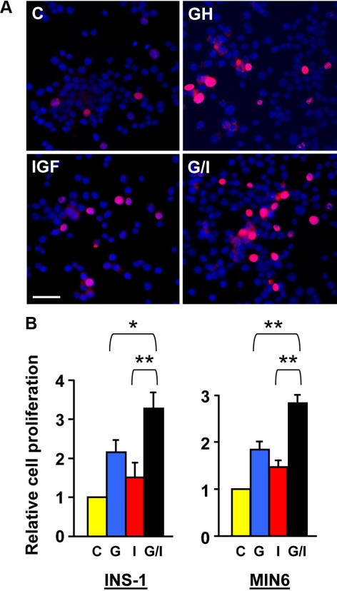
GH and IGF-I cotreatment augments β-cell growth. A, Representative images from BrdU incorporation assays. INS-1 cells were treated with vehicle (control), bGH (500 ng/ml), or IGF-I (20 ng/ml) or cotreated with bGH plus IGF-I (G/I) in serum-free medium for 24 h, in which BrdU was added in the last 4 h of treatment (chase phase). Cells with incorporated BrdU (proliferating cells) were detected by immunostaining with an anti-BrdU monoclonal antibody (red). Total cell numbers were measured by DAPI staining (blue). Scale bar, 50 μm. B, Cell proliferation rates determined by BrdU incorporation assays. The BrdU chasing periods in the INS-1 and MIN6 cells were 4 and 1.5 h, respectively. A total of approximately 1500 cells from 10 random imaging fields under each condition, as in A, were counted and the ratios of BrdU-positive cells (red) to the total cells (blue) (representing cell proliferation rates) were plotted with the control set as 1. Data are mean ± sem (n = 10). *, P < 0.05; **, P < 0.01. C, Control; G, GH; I, IGF-I.

