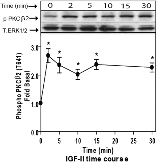
Fig. 4.
IGF-II induces activation of endogenous PKCβ2. Serum-starved HEK293 cells were stimulated with 10 nm IGF-II for the indicated times, and activation of PKCβ2 in whole-cell lysate samples was determined by immunoblotting with phosphorylation state-specific IgG. PKCβ2 phosphorylation is expressed as fold increase above the basal level in NS cells. A representative phospho-PKCβ2 and total ERK1/2 immunoblots are shown above a bar graph presenting mean ± sd of three independent experiments; *, P < 0.05 vs. nonstimulated (NS).

