Fig. 6.
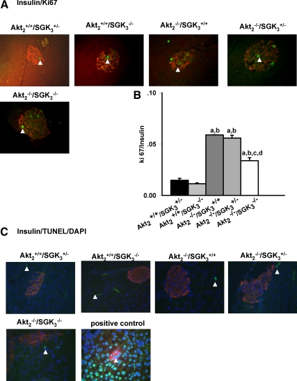
β-Cell proliferation and apoptosis in DKO mice. A, Pancreas sections from Akt2+/+/SGK3+/−, Akt2+/+/SGK3−/−, Akt2−/−/SGK3+/+, Akt2−/−/SGK3+/−, and Akt2−/−/SGK3−/− mice (7–9 wk of age; n = 3 per genotype) were stained with antibodies to insulin (red), Ki67 (green). Arrowheads indicate proliferating cells. B, The number of cells that were positive for both Ki67 and insulin were quantified as a percentage of total number of insulin-positive cells. At least 30 islets from three nonoverlapping pancreas sections were analyzed for each animal. C, Pancreas sections from above indicated genotypes were stained with TUNEL kit (green), antibodies to insulin (red), and 4′,6-diamidino-2-phenylindole (DAPI) (blue). Data are represented as mean ± sem. a, P < 0.05 vs. Akt2+/+/SGK3+/−; b, P < 0.05 vs. Akt2+/+/SGK3−/−; c, P < 0.05 vs. Akt2−/−/SGK3+/+; d, P < 0.05 vs. Akt2−/−/SGK3+/−.

