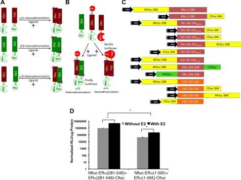
Fig. 1.
A and B, Scheme of independent and simultaneous split firefly and Renilla luciferase reporter protein complementation systems designed for studying ER-ligand induced homo- and heterodimerizations (ERα and ERβ) in cells, and noninvasively imaging in living animals by optical bioluminescence imaging. In this system, the LBD of ER (ERα and ERβ) are linked to the N- and C-terminal portions of luciferases, which then complement when the cells in which they are coexpressed are treated with ligands having different specificity and biocharacters for these receptors. The orientation (NH2 to COOH) of fusion proteins in different constructs were represented by labeling the different ends of the ER-LBD. C, Schematic diagram of different eukaryotic expression vectors constructed for the study in pcDNA (3.1) vector backbone. The schemes show the order of fragments of reporters (firefly luciferase: Fluc and Renilla luciferase: hRluc: N- and C- indicates the NH2 and COOH-terminal fragments of reporter proteins) and the ER (α and β) with respective sizes cloned to express different fusion proteins. D, The sensitivity of ER-dimerization system evaluated by vectors constructed to express fusion proteins of split-firefly luciferase fragments with either full-length or the LBD of ERα. HEK293T cells cotransfected with the vectors assayed for the complemented luciferase activity after exposure to ligand E2 (y-axis in log scale; *, P < 0.01). CMV, Cytomegalovirus.

