Figure 1.
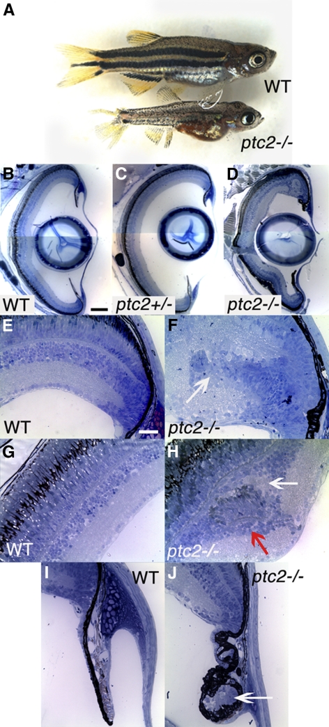
Juvenile ptc2−/− mutants possess peripheral retinal dysplasias and abnormalities at the ciliary zone. A small percentage of ptc2−/− mutants (∼2%) reach 6 weeks of age. ptc2−/− mutants are smaller than their wild-type siblings and have a disrupted pigmentation pattern (A). Histologic analyses of wild-type (B, E, G, I), ptc2+/− (C), and ptc2−/− (D, F, H, J) 6-week retinas. Retinal organization appeared normal in ptc2+/− mutants (C). In ptc2−/− mutants, retinal disorganization was found in the dorsal peripheral retina, whereas the rest of the retina was laminated normally (D). Within the dorsal peripheral retina of ptc2−/− mutants, ectopic neurons that appeared to be continuous with the CMZ and disrupted lamination were detected (F, arrow; n = 4/10). Ectopic neuronal clusters in the INL were also observed (H, n = 3/10). Neuronal clusters contained photoreceptor outer segments (H, red arrow) and were associated with an ectopic plexiform layer (H, white arrow). Morphologic abnormalities and ectopic cells (J, arrow) were observed in the ciliary zone (n = 3/10). Scale bar: (B–D) 100 μm; (E–J) 25 μm.

