Figure 5.
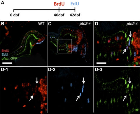
Continually proliferative cells in regions of retinal dysplasia in ptc2−/− mutants. A BrdU/EdU double-labeling experiment was performed on wild-type gfap:GFP (B) and ptc2−/−;gfap:GFP mutant retinas (C, D, D1–3). The fish were exposed to BrdU for 8 hours at 40 dpf, returned to their tanks for 2 days, injected with EdU at 42 dpf (6 weeks), and fixed for BrdU and EdU immunohistochemistry and detection (A). In wild-type retinas, cells that were proliferative at the time of fixation (EdU+) were mostly confined to the CMZ, whereas cells that were proliferative 2 days before fixation (BrdU+) had incorporated into the retina. In addition, a few GFP+ Müller glia were also either BrdU+ or EdU+ (B). In ptc2−/−;gfap:GFP retinas, in addition to BrdU+/GFP+ or EdU+/GFP+ Müller glia, BrdU+/EdU+ double-labeled cells were detected in the peripheral retina, and these did not express GFP (C, and white arrows in high magnification of boxed region in D and in D1–3, n = 3/3). Scale bars: (B, C) 100 μm; (D, D1–3) 20 μm.

