Figure 3.
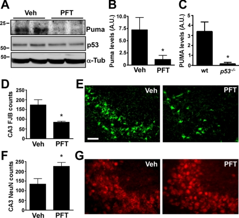
Puma induction after SE in mice is p53-dependent. A) Representative Western blots (n=1/lane) showing hippocampal Puma and p53 expression 8 h after SE in vehicle (Veh)- and pifithrin-α (PFT)-treated mice. Note that Puma levels are reduced in PFT-treated mice, while p53 levels were similar between groups. B) Graph showing significantly lower Puma levels in PFT-treated mice when compared to vehicle-treated mice after SE (n=3/group). C) Graph showing lower Puma levels in p53−/− mice when compared to wild-type (wt) mice 8 h after SE (n=3/group). D–G) Graphs (D, F) and representative photomicrographs (×40 lens; E, G) showing hippocampal damage 24 h after SE as assessed by counts of FJB-positive CA3 cells (D, E), and CA3 cells with normal-appearing NeuN immunoreactivity (F, G), in PFT-treated mice compared to vehicle-treated mice (n=4–7/group). Note significantly fewer FJB-positive cells and significantly more surviving neurons in PFT-treated mice. A.U., arbitrary units. *P < 0.05 vs. control. Scale bar = 100 μm.

