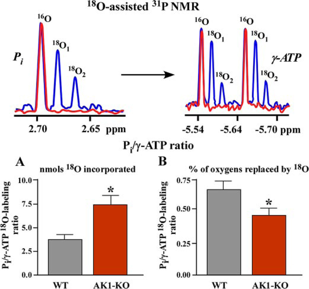
FIGURE 1. Disturbed intracellular energetic communication in AK1-KO hearts after ischemia-reperfusion assessed by 18O-assisted 31P NMR.
Upper panel, 18O-assisted 31P NMR recordings of 18O-induced shift (blue) in 31P NMR spectra of Pi (red) and γ-ATP (blue and red, respectively) indicating incorporation of one and two atoms of 18O and metabolic activity of these energetic intermediates. Lower panels, A and B, changes in Pi/γ-ATP labeling ratios (by 18O mass (A) and by percentage of 18O (B) incorporated into corresponding phosphoryls) in wild-type (WT) and adenylate kinase knock-out (AK1-KO) hearts after ischemia-reperfusion. Increased 18O mass in Pi compared with γ-ATP (A) along with diminished percentage labeling ratio (B) indicates disrupted communication between ATP consumption and ATP synthesis sites in AK1-KO hearts.

