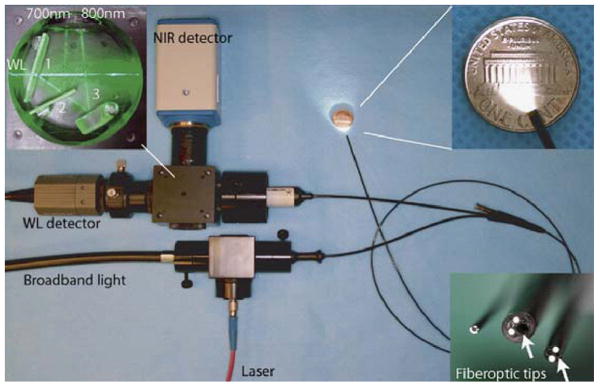
Fig. 1. Near-Infrared Small Animal Endoscope.
A modified fiber optic angioscope (1.4 mm diameter) collects both the white light and near-infrared (NIR) fluorescence images. The schematic for spectral processing is shown in the top left inset. The first dichroic mirror transmits white light from the endoscope and reflects the broad band near-infrared fluorescence. The second dichroic transmits and reflects fluorescence in the upper (750 to 800 nm) and lower (675 to 725 nm) NIR bands, respectively. The lower right inset shows several different angioscopes with diameters of 0.8, 1.6, and 2.4 mm.

