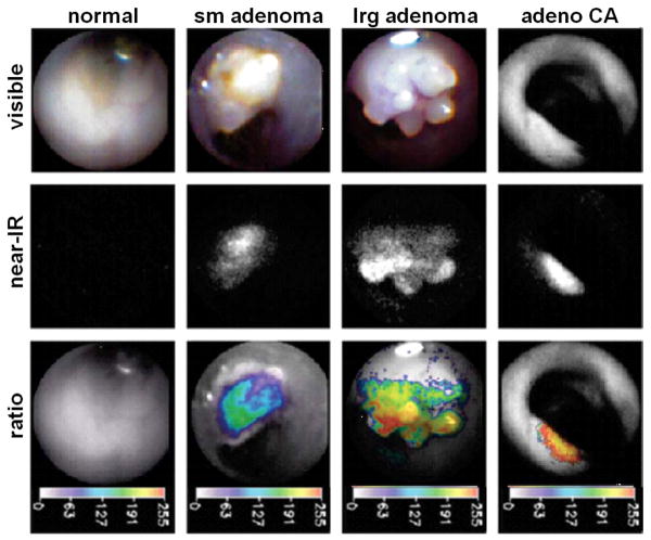
Fig. 3. Endoscopic images from APCmin/+ mouse.
In the first row, in vivo white light images of normal mucosa, small adenoma, large adenoma, and adenocarcinoma collected with a 1.4 mm diameter angioscope in the colon of APCmin/+ mice are shown. In the second row, NIR images from the same mucosal regions are collected after i.v. injection of the protease-activatable probe and reveal increased fluorescence intensity. In the third row, the ratio of the NIR images collected after injection of protease-activatable and non-activatable probes are shown in pseudocolor. This ratio corrects for differences in object distance, collection angle, tissue reflectance and probe delivery. Increased intensity on the ratio images was found to correlated to cathepsin B expression on immunohistochemistry.

