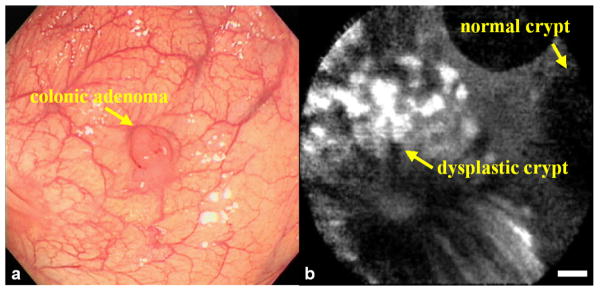
Fig 6. Targeted microscopic imaging in vivo.
a) Conventional white light endoscopic image of colonic adenoma, and b) in vivo confocal fluorescence image collected after topical administration of FITC-labeled target peptide shows preferential binding to dysplastic colonocytes. The dysplasia:normal border shows an average target-to-background ratio of 21, scale bars 20 μm.

