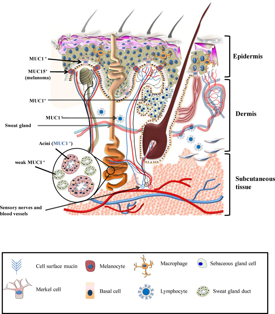
Figure 2. Schematic diagram of the normal skin showing an expression of various mucins.
The normal skin is comprised of three layers, the epidermis (most superficial), the dermis and the subcutaneous tissue (innnermost layer). MUC1, MUC2, MUC5AC and MUC6 are not expressed by the normal epidermis. MUC4 is weakly expressed by the epidermis in a proportion of tissue sections. The luminal surfaces of eccrine and apocrine glands and the degenerate cells comprising the secretions of the sebaceous glands are positive for MUC1. In apocrine glands (e.g. Moll’s glands of the eyelids), MUC1 is strongly expressed on the cell membrane and weakly in the cytoplasm of active glandular elements. MUC1 staining is also observed in the secretions present in the lumen of these glands. The acini of both sebaceous and sweat glands express MUC1 but not MUC2, MUC6 or MUC5AC (Except Bartholin’s glands which express MUC5AC). Ducts of these glands, however, are either negative (using antibodies specific for underglycosylated MUC1) or weakly positive (using antibodies that recognize MUC1 independent of its glycosylation status) for MUC1. Merkels cells express an under-glycosylated form of MUC1. Lymphocytes present in the normal skin express an under-glycosylated form of MUC1. Melanocytes normally do not express mucins but MUC15 mRNA expression was shown to be aberrantly upregulated in thin melanomas.

