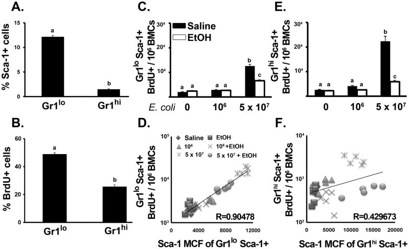
Figure 4.
Marrow granulocyte proliferation. Percent of (A) Sca-1+ and (B) BrdU+ cells in marrow Gr1+ populations from control mice. Number of BrdU+ cells in (C) Gr1loSca-1+ and (E) Gr1hiSca-1+ populations after 24 h E.coli (5 × 10⁷) challenge. Correlation between Sca-1 MCF and BrdU+ cell number in (D) Gr1loSca-1+ and (F) Gr1hiSca-1+ cells. Data are mean ± SEM (N=5). Bars with different letters are statistically different (p < 0.05).

