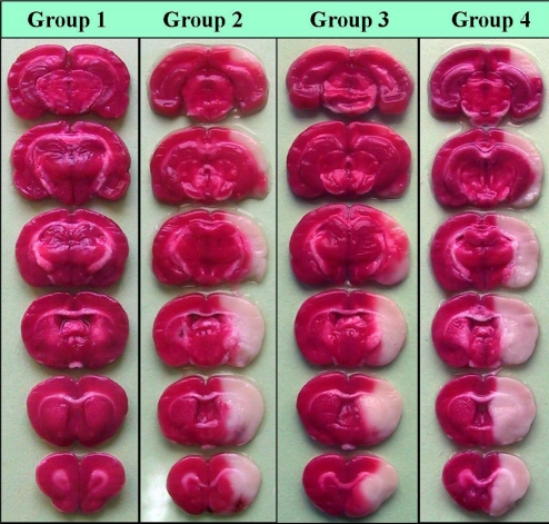
Figure 3.
Photographs illustrating the coronal sections of rat brain slices stained with TTC 24 hrs after neck surgery in sham (group 1) or after 60 min occlusion of middle cerebral artery and 24 hrs reperfusion in control ischemic (group 2), and enalapril pre-treated ischemic rats (0.03 or 0.1 mg/kg, groups 3 and 4) as described in the text. MCA occlusion induced different magnitudes of infarctions in the right hemispheres without affecting the left sides. Non-ischemic areas are colored deep red, whereas, ischemic areas are white. Note the similarity of ischemic areas of brain slices of groups 2 and 4. There is a marked decrease in the ischemic areas of group 3.

