Figure 2.
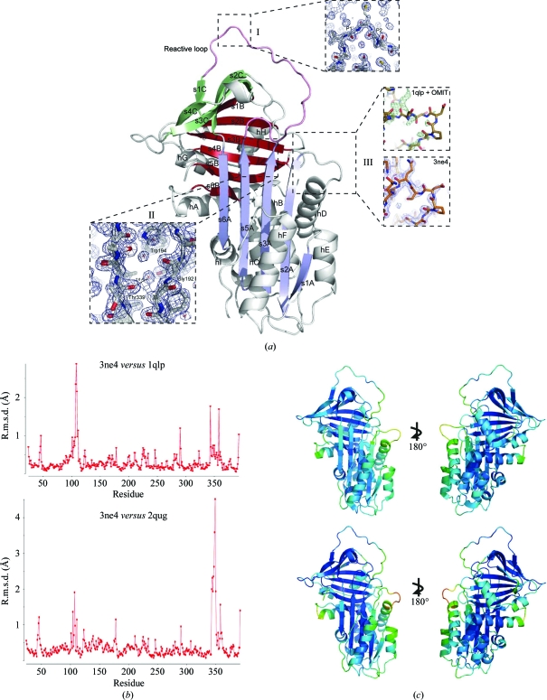
(a) 1.8 Å resolution crystal structure of α1-antitrypsin (PDB entry 3ne4) with α-helices and β-strands labelled (e.g. helix A, hA; strand 1 of β-sheet A, s1A). Strands within a β-sheet are colour-coded together (A, blue; B, bronze; C, green). Detail is shown for the following. Box I, the reactive centre of the molecule in the canonical conformation. Box II, the ‘breach’ position that is the site of initial intramolecular loop insertion during monomeric conformational transitions. Box III, the fit of the hD–s2A turn. The upper panel shows the rigid fit of 1qlp (gold) together with the initial OMIT map (F o − F c at 3σ density when residues 105–110 are omitted; positive difference density in green, negative in red). The lower panel shows the final fit of 3ne4 (orange) to the final map (blue, 2F o − F c at 1σ density). (b) R.m.s.d. for observed α1-antitrypsin residues in 3ne4 compared with 1qlp (upper panel) and 2qug (lower panel) calculated using the SUPERPOSE program from the CCP4 suite (Winn et al., 2011 ▶). (c) Comparison of B factors in 1qlp (above) and 3ne4 (below). Low/high values are indicated by rainbow-spectrum colouring by PyMOL using a preset scale (blue for low to red for high). Whilst overall B factors are lower in 3ne4 (range 9.60–83.99 Å2, mean 23.9 Å2) than 1qlp (range 13.82–96.92 Å2, mean 38.4 Å2), the hD–s2A turn is associated with increased values in both relative to the global values. Other regions that show relative increases in B factor are the C-terminal end of helix A and the upper turn of helix F, which is believed to be dynamic in solution and to remodel during formation of the intermediate.

