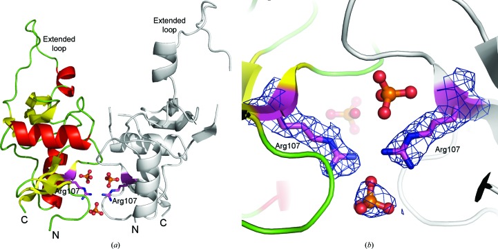
Figure 1.
Overall structure of the H107R variant of mouse NKR-P1A and a detailed view of the interface between monomers. (a) Chain A is represented by coloured secondary-structure elements, chain B is shown in grey, phosphate ions are shown in ball-and-stick representation and side chains of Arg107 are represented by sticks. (b) A detailed view of the interface between the monomers and the binding of the additional phosphate ion. The 2F o − F c map around the phosphate and Arg107 is contoured at the 1σ level (blue).

