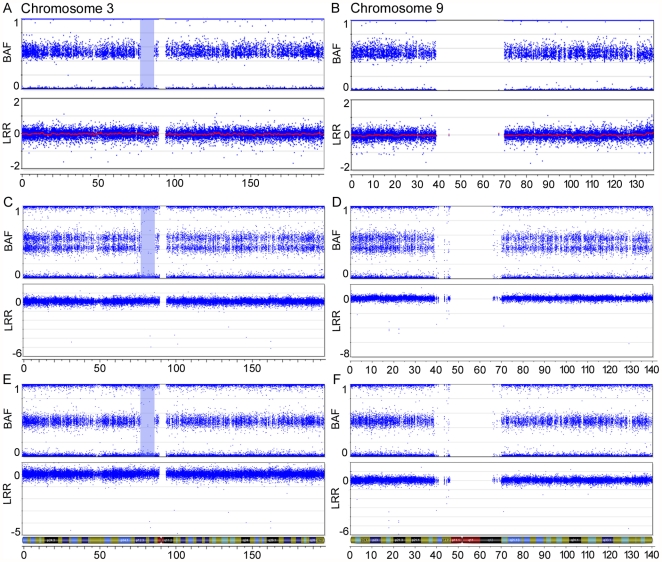
Figure 1. SNP array imaging results for chr3 and chr9 of the benign serous tumour 1781T.
SNP array imaging results for chr3 (A, C, E) and chr9 (B, D, F) of the benign serous tumour 1781T, using Illumina's HumanHap300-Duo Genotyping BeadChip (A and B) and Illumina's Human610-Quad Genotyping BeadChip (C–F). Two different DNA preparations were used with the HumanHap610-Quad Genotyping BeadChip. The top plot of each figure shows the B allele frequency (BAF) for each SNP marker aligned to its chromosomal position. In heterozygous diploid cells, alleles are present in AA, AB or BB pairs. The B Allele frequencies for these possible allele pairs are 0, 0.5 or 1, respectively. Any deviation from this ratio indicates a chromosomal aberration. In one DNA preparation, the double row in the BAF plot indicates allelic imbalance of SNP markers across the entire chromosome (C and D). A 9.1 Mb ROH is observed on chr3 and is highlighted in blue. No markers are located in the centromeric region of either chromosome, as noted by a lack of markers in both the B allele frequency and Log R ratio (LRR) plots. The bottom plot of each figure contains the Log R ratio, which provides an indication of the copy number for each SNP marker aligned to its chromosomal position. Note the absence of a drop in the Log R ratio in the highlighted ROH.

