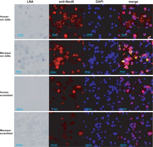
Figure 6. In situ staining of miR-320b in prefrontal cortex.
First row: Human newborn prefrontal cortex sections were hybridized with miR-320b LNA-probes (far left); anti-NeuN antibodies staining neuron nuclei (center left) and DAPI staining DNA (center right); and a merged image showing LNA probes in yellow (far right). The LNA picture was taken under bright field at 40× magnification, and the DAPI and anti-NeuN pictures used the fluorescent channel. For the merged image the LNA signal was modified to a green color scale. Second row: Rhesus macaque prefrontal cortex sections processed and displayed in the same way as human. Third and fourth rows: Human and rhesus macaque sections were treated the same way as described, but using LNA probes with scrambled miRNA sequences as a negative control. The scale bar indicates 50.0 µm. See Materials and Methods for details.

