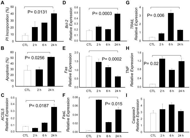
Figure 3. Effect of PMA+Io activation in Jurkat T cells.
Jurkat T cells were cultured in the presence or absence of PMA+Io for up to 24 h. A) Cells were collected at defined times, washed with PBS, fixed with 70% Ethanol, stained with Propidium Iodide (PI) and analyzed in a FACSCaliburTM flow cytometer to determine the apoptotic hipodiploid cell fragments (defined as percentage of PI incorporation). B) Cells were washed twice with PBS and double stained with Annexin V and PI, then analyzed by FACS to determine the percentage of apoptotic cells (Annexin V positive and double positive cells). C–I) Total RNA was extracted, cDNA synthesized and qRT-PCR implemented to determine mRNA expression. Results are given by means of three independent experiments and the bars show the standard deviation. P-value has been calculated with the paired Student t test.

