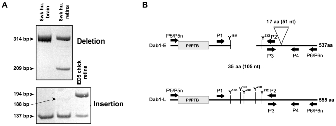
Figure 1. RT-PCR analysis of Dab1 deletion and insertion regions.
(A) cDNAs synthesized from poly(A)+ RNA from human fetal brain (8 wks gestation), retina (8 wks gestation) and chick retina (E5) were amplified using P1 and P2 primers for deletion analysis and P3 and P4 primers for insertion analysis. Sizes of amplified bands are indicated. (B) Schematic representation of human Dab1-E and Dab1-L proteins and relative positions of primers used for RT-PCR amplification.

