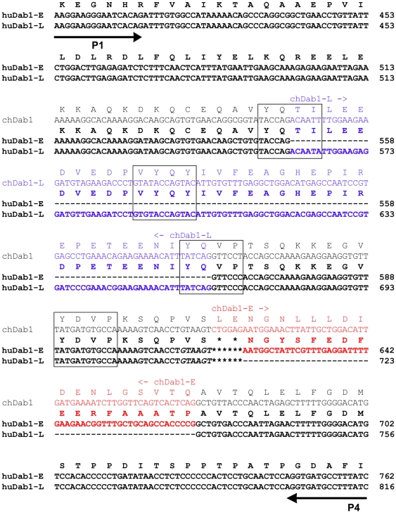
Figure 2. Sequence alignment of P1/P4-amplified Dab1 fragments.
HuDab1-E, huDab1-L and chDab1 nucleotide and amino acid sequences are as indicated. The Dab1-L-specific 105 nt two-exon (7/8) region is indicated in blue while the huDab1-E-specific 51 nt exon 9B (57 nt in the case of chDab1-E) is indicated in red. SFK (Y185 and Y198) and Abl/Crk (Y220 and Y232) recognition motifs are boxed. The six italicized nucleotides were only present in a subset of the products analysed.

