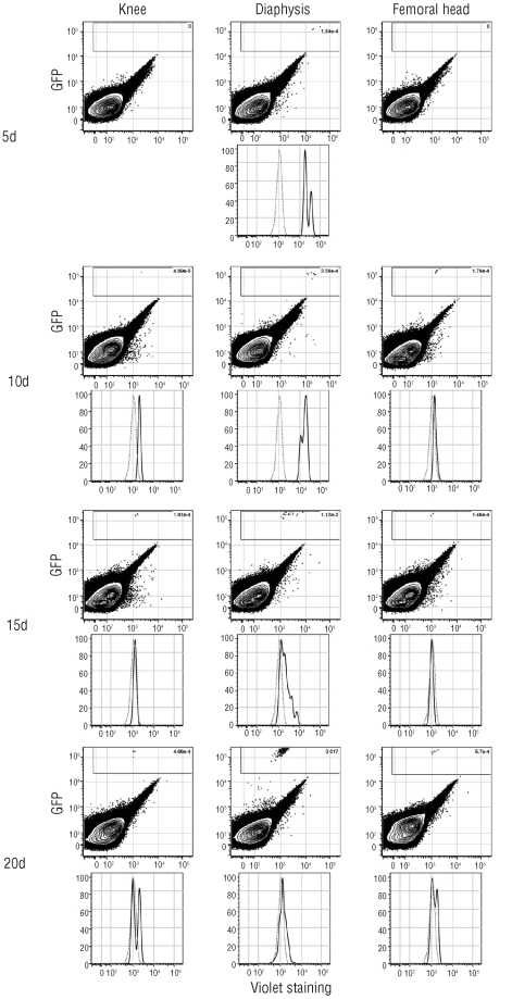
Figure 3.
Kinetics of proliferation of engrafted MLL-ENL leukemic cells after CFSE labeling. Flow cytometric analysis of CellTrace violet labeled GFP+ leukemic cells detected in knee, diaphysis and femoral head of non-conditioned 129/Sv mice injected with 1,000 CellTrace violet labeled GFP+ MLL-ENL leukemic cells. Upper panels: FACS analyses of GFP and violet fluorescence 5, 10, 15 and 20 days after injection. Lower panels: mean fluorescence of violet labeled GFP+ leukemic cells (black line) or unlabeled GFP+ leukemic cells (dash line) 5, 10, 15 and 20 days after injection. The data are representative of 3 experiments with 5 mice per day of acquisition.

