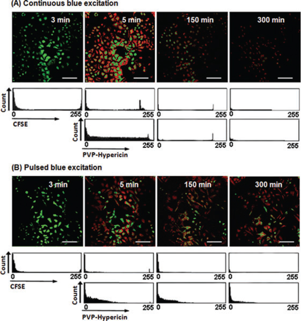
Fig. 7.
A5-h imaging of the dual-marked A549 cells with CFSE and PVP-Hypericin. Images were collected with a 20× objective (air for phase contrast). (A) Fluorescence signals and cell proliferation under continuous blue excitation. (B) Fluorescence signals and cell proliferation under pulsed blue excitation. The scale bars represent 50 µm.

