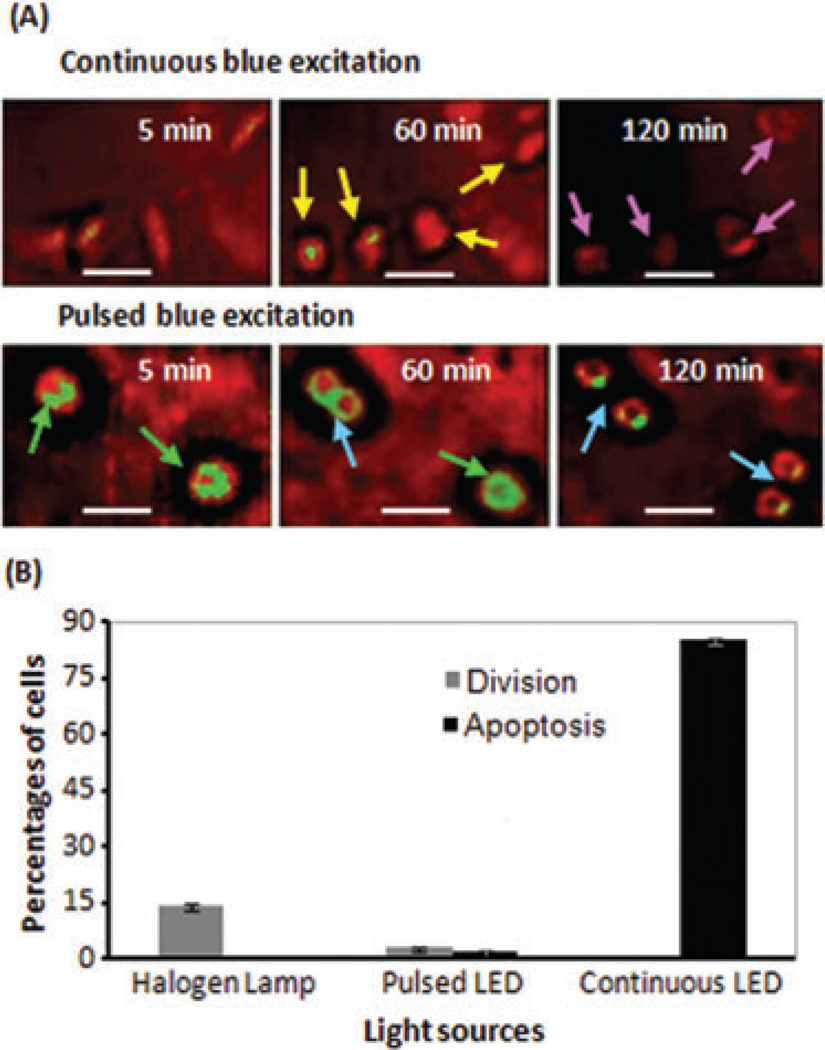
Fig. 8.
(A) Monitoring of apoptotic and divided A549 cells, dual-marked with CFSE and PVP-Hypericin, under continuous and pulsed excitation. Cells excited with continuous blue LED round up (yellow arrows) and after 120 min most cells are apoptotic (pink arrows). By applying pulsed blue LED the photodamages are delayed and even dynamic monitoring of cell division (blue arrows) is possible. (B) Comparison of the percentages of division and apoptosis in dual-marked cells under blue-LED excitation with nonmarked reference cells under the halogen lamp, during 5-h imaging. Error bars represent the standard deviation. The scale bars represent 10 µm.

